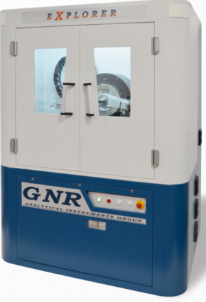
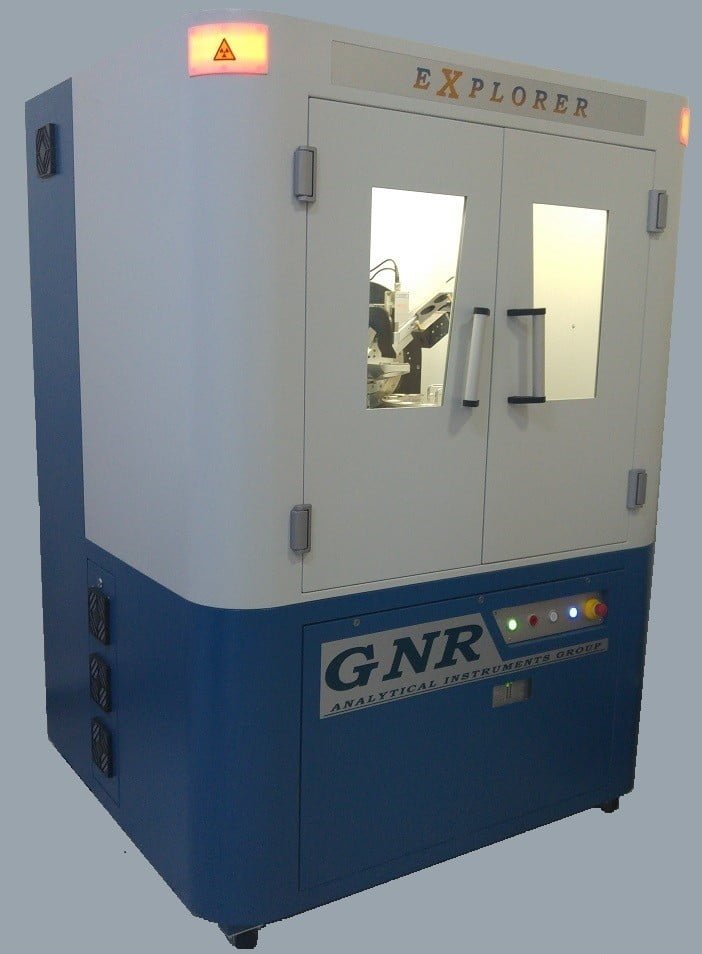
Difractometru cu raze X - GNR EXPLORER
X-Ray Diffraction

X-Ray Diffraction (XRD) is a non-destructive technique for the qualitative and quantitative analysis of the crystalline materials, in form of powder or solid.
GNR has developed, in cooperation with academic and industrial users, a set of technically advanced and flexible diffractometers able to satisfy different level of requirements and different operating budget.
GNR XRD Product Portfolio covers a huge range of applications for materials characterization and quality control of crystalline or non-crystalline materials such as powders, specimens, thin films or liquids.

Basically XRD is obtained as the „reflection“ of an X-ray beam from a family of parallel and equally spaced atomic planes, following the Bragg’s law: when a monochromatic X-ray beam with wavelength l is incident on lattice planes with an angle q, diffraction occurs if the path of rays reflected by successive planes (with distance d) is a multiple of the wavelength.
Qualitative analysis (phase analysis) can be done thanks to the comparison of the diffractogram obtained from the specimen with a huge number of patterns included in the official databases. Single phases and/or mixtures of phases can be analysed with the programs available today.
Many Investigations can be performed with the help of X-ray diffraction.
Residual Stress Forces that results in a small compression or dilatation of the d-spacing. With XRD it is possible to measure the strain (the deformation of the original lattice) and the stress is calculated thanks to the knowledge of the elastic constants of the material
Texture It is the preferred orientation of the crystallites in a specimen. If a texture in a material is present, the intensity of a diffraction line changes with the orientation of the sample respect to the incident beam.
Crystallite size and micro strain These information are obtained by the analysis of the width and the shape of the diffraction lines.
Structure analysis XRD is used to investigate the crystallographic structure of a material. The position and the relative intensities of the diffraction lines can be correlated to the position of the atoms in the unit cell, and its dimensions. Indexing, structure refinement and simulation can be obtained with specific computer programs.
Thin film Keeping the incident beam at low angles, it is possible to investigate the properties of multilayers, minimising the interference of the substrate. On the same way, reflectometry can be performed.
Explorer is a multi purpose – Theta/Theta – high resolution diffractometer which, thanks to its direct drive torque motors, offers top performances in many analytical areas, from phase analysis to determination of microstructural properties on bulk or thin film materials.
Thanks to its modularity and the wide range of accessories and attachments available, EXPLORER allows to perform measurement in different configurations: traditional X-Ray Powder Diffraction (XPD), Reflectometry (XRR), Grazing Incidence X-Ray Diffraction (GIXRD), High Resolution X-Ray Diffraction (HRXRD), Total X-Ray Fluorescence (TXRF), Residual Stress and Texture X-Ray Diffraction.
The modularity and the flexibility of the GNR X-Ray Explorer allows to start with an entry-level system which can be upgraded to meet new requirements. GNR could supply a wide range of X-ray sources, optics, sample holders, detectors and configurations to satisfy all the analytical needs.
With no limits to its applications, Explorer modular system offers high performances in all analytical areas, ranging from phases quantification of mixtures, to the determination of microstructural properties as residual stress and preferred orientation of crystallites on bulk materials as well as on thin films.
Applications:
- Routine Cystalline phase identification and quantification
- Crystallite size – lattice strain and crystallinity calculation
- Polymorph screening and crystal structure analysis
- Residual Stress and Retained Austenite Quantification
- Thin Films, Depth Profiling and non ambient analysis
- Phase Transition monitorin, tecture and preferred orientations
The optics permit switches between Bragg-Brentano, focusing and parallel beam geometry using Johansson or parabolic mirror monochromators.
The high resolution reflectometry studies can be performed with EXPLORER to characterise layer thickness (from 1 to 500 nm with an accuracy better than 1%), density (with an accuracy better than ± 0.03 g/cm3), surface and interface roughness (from 0 to 5 nm with an accuracy better than ± 0.1 nm).
Measurements at low angles and a thin film attachment for parallel beam geometry allow the study of thin films and multilayers.
The coupling between a parabolic mirror monochromator and a channel-cut crystal mounted on the incident beam allows to realise a monochromatic parallel beam with high intensity and low divergence, suitable for high resolution measurements.
| X Ray Generator | Maximum Output Power | 3 kW (option: 4 kW) |
| Output Stability | < 0.01 % (for 10% power supply fluctuation) | |
| Max Output Voltage | 60 kV | |
| Max Output Current | 60 mA (option: 80 mA) | |
| Voltage Step Width | 0.1 kV | |
| Current Step Width | 0.1 mA | |
| Ripple | 0.03% rms < 1kHz, 0.75% rms > 1kHz | |
| Preheat and Ramp | Automatic preheat and ramp control circuit | |
| Input Voltage | 220 Vac +/-10%, 50 or 60 Hz, single phase | |
| Size | Width 48.3 cm, height 13.3 cm, depth 56 cm | |
| X-Ray Tube | Type | Glass (option: Ceramic), Cu Anode, Fine Focus (options: any kind of X-Ray tube) |
| Focus | 0.4 x 8 mm FF (other options available) | |
| Max Output | 3.0 kW | |
| Goniometer | Configurations | Horizontal and vertical Theta/2Theta and Theta/Theta geometry |
| Measuring circle diameters | 400 – 500 – 600 mm or any intermediate settings | |
| Scanning Angular Range | – 110° < 2 Theta < + 168° (according to accessories) | |
| Smallest selectable stepsize | 0.0001° | |
| Angular reproducibility | +/- 0.0001° | |
| Modes of operation | Continuous scan, step scan, theta or 2 theta scan, fast scan, theta axis oscillation | |
| Divergence slits | 4°; 2°; 1°; 1/2°; 1/4° | |
| Anti-Divergence slits | 4°; 2°; 1°; 1/2°; 1/4° | |
| Receiver slits | 0.3; 0.2; 0.1 mm | |
| Soller slits | 2° | |
| Detector | Type | Scintillation counter Nal (options: YAP(Ce); multistrip) |
| Countrate | 2 x 10(6) cps | |
| HV/PHA | High voltage supply 600 – 2000 V, gain, low, central and high level control | |
| Case | Dimensions | Width 1400 mm, heigh 1800 mm, depth 850 mm |
| Leakage X-rays | < 1 mSv/Year (full safety shielding according to the international guidelines) | |
| Processing Unit | Computer Type | Personal Computer, the latest version |
| Items controlled | X-ray generator, goniometer, sample holder, detector, counting chain | |
| Basic Data Processing | Qualitative and quantitative phase analysis. Rietveld analysis, crystalline structural analysis, crystallite size and lattice strain, crystallinity calculation, strain, reflectometry. |
Software
Data Collection Programs
GNR offers a large variety of acquisition programs, for standard as well as for customized hardware configurations. the list includes programs for powder and high resolution diffractometers, retained austenite, data acquisition of stress (plane and triaxial) and thin films (XRD and GIXRD).
SAX
Single peak analysis; peak treatment. Background subtraction, smoothing, deconvolution and peak localisation. Structural Analysis, Crystallite Size, Lattice Strain, Reflectometry, Quantitative Analysis.
Search and Match: MATCH!
Rietveld refinement, Display and compare multiple diffraction partners, Directly view specific phases/entries, instant usage of additional information, saving of selection criteria, Comfortable definition of background, Improved zooming facilities, Batch processing and Automatics.

